- Mon - Sat 10:00 - 07:30
- PAN Empire, Opp. Silver Heights, Rajkot, Gujarat 360005
- Mon - Sat 10:00 - 19:30
- One World, Rajkot
- +91 962-484-9978
- Mon - Sat 10:00 - 19:30
- One World, Rajko 360007
- +91 962-484-9978
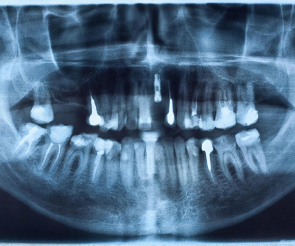
Accurate diagnosis begins with our advanced OPG & CT Scan providing precise imaging for optimal treatment planning.
Clear, detailed imaging for precise dental diagnostics.
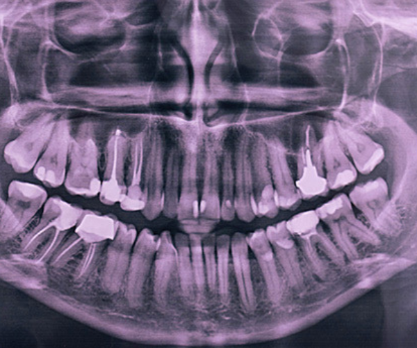
1.What is an OPG?
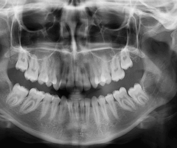
Description: An OPG is a panoramic X-ray that captures a comprehensive view of the upper and lower jaw, including the teeth, bones, and surrounding tissues, in a single image.
Purpose: It provides a broad overview of the dental and skeletal structure, allowing for the assessment of various conditions.
2.Uses of OPG
Assessment of Dental Health: Helps in evaluating the health of teeth, detecting cavities, and identifying problems with tooth roots.
Orthodontic Planning: Essential for planning orthodontic treatments, including braces and other corrective procedures.
Evaluation of Jaw and Bone Structure: Assists in diagnosing issues such as jaw disorders, bone abnormalities, and impacted teeth.
Detection of Pathologies: Useful for identifying cysts, tumors, and other anomalies within the oral and maxillofacial region.
3.Procedure
Preparation: No special preparation is required. Patients are asked to remove any metal objects (e.g., earrings) that might interfere with the image.
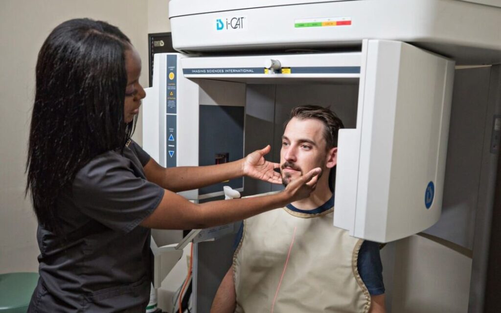
Process: The patient stands or sits in a stationary position while the X-ray machine rotates around their head, capturing the panoramic image.
4.Benefits
Comprehensive View: Provides a complete view of the dental and skeletal structures in one image.
Quick and Non-Invasive: The procedure is fast, non-invasive, and involves minimal radiation exposure.
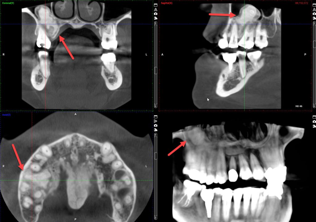
1.What is a CT Scan?
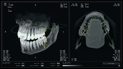
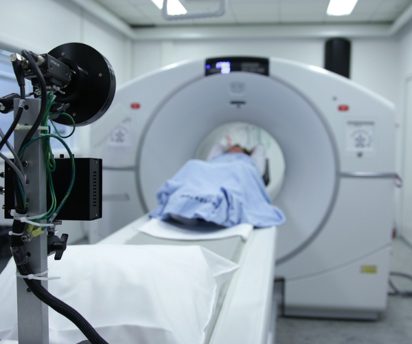
Description: A CT scan uses X-rays and computer technology to create detailed cross-sectional images (slices) of the body, including the oral and maxillofacial regions. It offers a more detailed view compared to standard X-rays.
Purpose: Provides in-depth information about the structure and composition of tissues and bones.
2.Uses of CT Scan
Detailed Assessment: Allows for a detailed examination of complex dental and maxillofacial conditions, including the evaluation of bone density and the presence of pathology.
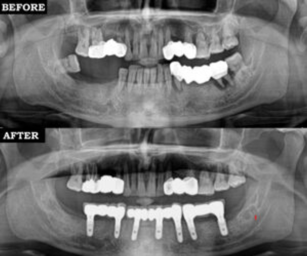
Pre-Surgical Planning: Critical for planning complex dental and surgical procedures, including dental implants and reconstructive surgery.
Trauma Evaluation: Assists in assessing injuries to the facial bones and other critical structures in cases of trauma.
Diagnosis of Diseases: Helps in diagnosing diseases affecting the sinuses, jaw joints, and other areas within the oral and maxillofacial region.
3.Procedure
Preparation: Patients may be asked to remove metal objects and, in some cases, to follow specific dietary or fluid intake instructions.
Process: The patient lies on a table that slides into a CT scanner. The scanner takes multiple X-ray images from different angles, which are then processed by a computer to create detailed cross-sectional images.
4.Benefits
High-Resolution Images: Provides highly detailed images that are crucial for accurate diagnosis and treatment planning.
Versatility: Can be used to examine various structures and conditions within the oral and maxillofacial regions.
State-of-the-Art Equipment: Utilizes advanced imaging technology to ensure high-quality, accurate results.
Expert Interpretation: Images are reviewed and interpreted by experienced radiologists and specialists.
Comprehensive Care: Integrates imaging results into a holistic treatment approach, ensuring precise diagnosis and effective treatment planning.
Patient Comfort: Focuses on providing a comfortable and efficient imaging experience.
Take the First Step Toward a Healthier Smile—Book Your Appointment Today at Smile Plus Hospital!
Smile Plus Hospital is where advanced dental care meets modern aesthetics — a trusted space in Rajkot dedicated to creating confident smiles and enhancing natural beauty with precision, compassion, and expertise.